
The following blog post is for entertainment and informational purposes only. It is not intended to provide medical advice or diagnosis. Please consult your doctor before making any health-related decisions.
Microglia function as the brain's resident immune cells. They scan neural tissue continuously, clearing debris, regulating inflammation, and pruning the synaptic connections that shape memory and learning. This cellular work underlies every aspect of long-term cognitive performance, from active mental engagement that research links to brain health to the daily baseline upkeep that keeps neurons alive across decades. When microglia malfunction, the result is chronic neuroinflammation. That inflammation connects directly to Alzheimer's disease, Parkinson's disease, and multiple sclerosis.
What do microglia do in the brain?
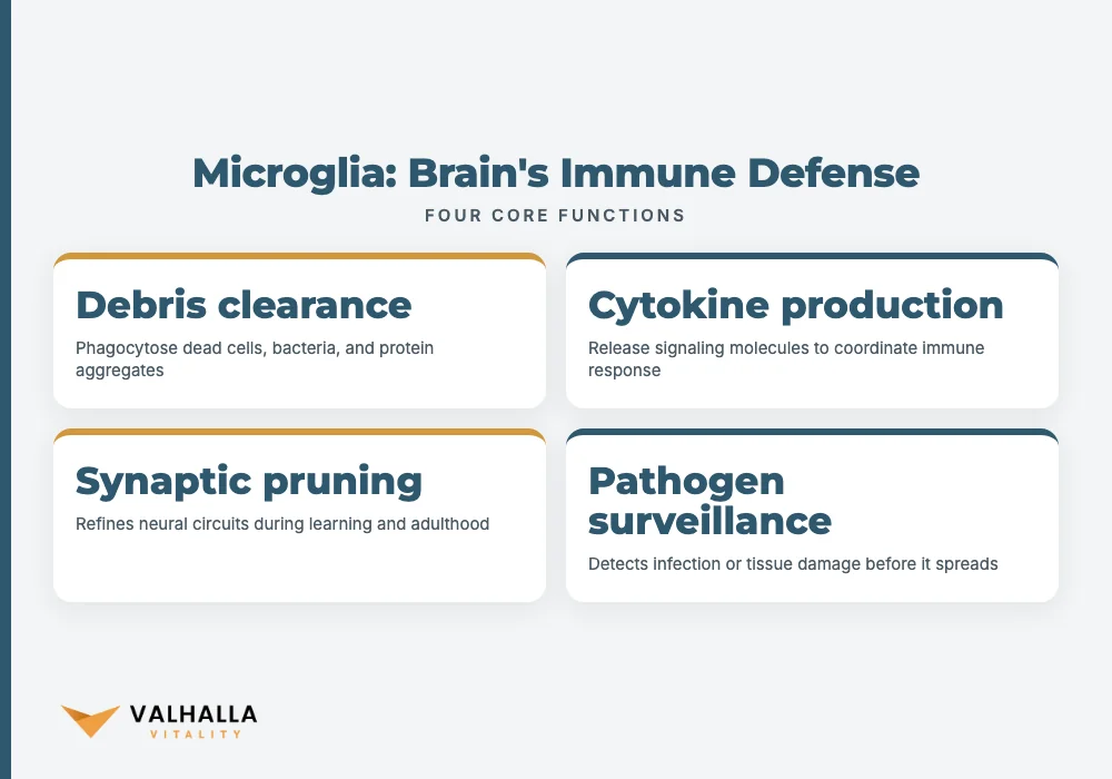
Microglia are the brain immune cells that form the central nervous system's dedicated immune defense. They work every moment of every day. A 2025 paper by Hume in Trends in Neurosciences makes the stakes concrete: neural tissue without functioning microglia develops chronic infection, progressive neurodegeneration, and uncontrolled toxic accumulation. Microglia function is not optional maintenance. It is the structural baseline on which healthy neural tissue depends.
Their work organizes into four overlapping categories:
| Core function | How microglia execute it | Why it matters |
|---|---|---|
| Debris clearance | Phagocytose dead cells, bacteria, and protein aggregates | Prevents neurotoxic buildup that disrupts neural signaling |
| Cytokine production | Release pro- and anti-inflammatory signaling molecules | Coordinates the immune response and its resolution |
| Synaptic pruning | Address weak or redundant synaptic connections | Refines neural circuits during learning and throughout adulthood |
| Pathogen surveillance | Continuously sample the extracellular environment | Detects infection or tissue damage before it spreads |
Microglia function at the cellular level
Each aspect of microglia function operates in close coordination with the others: debris clearance prevents the toxic signals that would otherwise sustain chronic cytokine release, and balanced cytokine output in turn supports appropriate synaptic pruning.
Phagocytosis is the most visible of these functions. When a neuron dies or a protein aggregate forms, microglia engulf and break it down before damage spreads. Left unchecked, this debris accumulates into the toxic clusters central to Alzheimer's pathology.
Cytokine production matters equally. Microglia release pro-inflammatory signals when they detect damage. Once the threat resolves, they shift to anti-inflammatory cytokines. This two-phase switch defines a healthy inflammatory response. When it fails, inflammation persists indefinitely.
Synaptic pruning adds a third dimension. Microglia shape neural circuits throughout adulthood, not only during development. This is why microglial function affects everyday memory consolidation and cognitive flexibility, not just post-injury repair.
Beyond these core tasks, microglia release neurotrophic factors that support neuron survival and help maintain the blood-brain barrier. A 2021 study by Wang et al. in Neural Plasticity examined how epigenetic modifications shape microglial behavior across neurodegenerative diseases, showing that the same cellular population can behave quite differently depending on the molecular context in which it operates. Microglia are not a uniform tissue. They are a finely calibrated system that adapts to local demands throughout the brain.
Microglia function and activation states
Microglia do not occupy a single fixed mode. Their phenotype spans a continuous spectrum, shifting in response to what they detect in the neural environment.
In a healthy brain, microglia are ramified, extending long branching processes that survey surrounding tissue. A 2024 study by Kou et al. in Ageing Research Reviews found that microglia follow a circadian rhythm, altering their inflammatory output across the day/night cycle. Disrupting that rhythm through chronic sleep deprivation or irregular schedules shifts baseline microglial activity toward inflammation, even without direct neural injury.
When damage is detected, microglia shift to an activated phenotype. They retract their processes, concentrate their cell bodies, and mount a response. Activation is not inherently harmful. It is the appropriate reaction to an acute threat. The problem begins when activation becomes chronic.
A 2025 review by Valiukas et al. in The Journal of Prevention of Alzheimer's Disease identifies multiple distinct microglial activation states, each with a different functional profile. Some promote tissue repair. Others amplify damage. Whether a response is protective or destructive depends on the duration and intensity of the trigger, and on whether the inflammatory off-switch functions normally.
Several factors push microglia into sustained activation beyond a proportionate response. Age-related cellular changes reduce the efficiency of the off-switch. Accumulated protein aggregates provide a chronic stimulus that microglia cannot fully clear. Metabolic dysfunction alters the signaling environment in ways that keep microglia sensitized. Each factor compounds the others over time. This compounding pattern is why maintaining healthy microglia function across the lifespan requires attention to multiple intersecting risk factors rather than any single variable.
What happens when microglia become overactive?
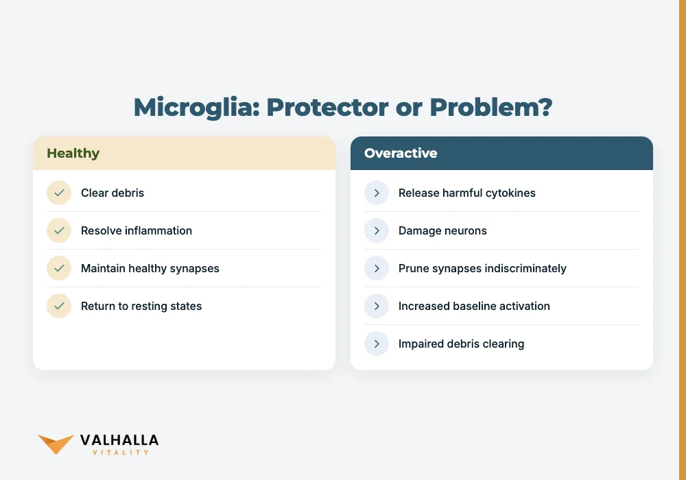
Chronically overactive microglia shift from protectors to drivers of neural damage. Rather than clearing debris and resolving inflammation, they release pro-inflammatory cytokines and reactive oxygen species in a sustained pattern that harms the neurons they are supposed to defend.
Common brain inflammation causes that prime microglia toward this state include:
- Traumatic brain injury, including subconcussive events
- Metabolic conditions such as obesity, insulin resistance, and cardiovascular disease
- Chronic systemic infection or persistent inflammatory signaling
- Normal aging and the cellular senescence that accumulates with it
- Chronic sleep disruption and circadian dysregulation
Microglia in aging brains show increased baseline activation, a reduced ability to return to resting states, and impaired phagocytic efficiency. The result is a chronic, low-grade neuroinflammatory environment that compounds tissue damage year over year.
Overactivation also disrupts synaptic pruning. Hyperactive microglia prune synapses indiscriminately, removing connections that should be maintained. This excessive pruning appears in models of early Alzheimer's disease, where synaptic loss precedes overt neurodegeneration by years.
Peripheral inflammation is a significant driver. Conditions like obesity, cardiovascular disease, and chronic infection generate systemic inflammatory signals that cross into the central nervous system. A 2025 review by Shi et al. in Science shows that peripheral immune signals reliably alter microglial behavior even when the blood-brain barrier remains largely intact. What happens in the body reaches the brain. For this reason, systemic inflammatory conditions are now understood as direct modulators of microglia function, not merely indirect contributors to cognitive risk.
What diseases are linked to dysfunctional microglia?
Every major neurodegenerative condition involves significant microglial dysfunction. The mechanism differs by disease, but the shared pattern is a microglial population that can no longer respond proportionally and stand down.
Alzheimer's disease is the most studied case. Microglia cluster around amyloid plaques in an initial attempt to clear them. Over time, this clearing effort fails and microglia shift into states that amplify neuroinflammation rather than resolve it. The 2025 review by Valiukas et al. identifies specific activation states that actively drive synaptic loss in Alzheimer's disease brains, confirming that microglia are mechanistic contributors to disease progression rather than passive bystanders.
Parkinson's disease involves sustained microglial activation in the substantia nigra, where dopamine-producing neurons degenerate. The resulting neuroinflammation accelerates neuron loss and worsens motor symptoms over time. Kou et al. (2024) identify microglial circadian regulation as a potential therapeutic target, raising the possibility that timing-based interventions may interrupt this inflammatory cycle.
Multiple sclerosis combines autoimmune and microglial mechanisms. Microglia can occupy both protective and damaging activation states in the MS brain, sometimes simultaneously in different lesion zones. Selectively amplifying repair-oriented microglial states, rather than applying broad immune suppression, has become a research priority.
Ischemic stroke shows both sides of microglial activity with unusual clarity. A 2018 study by Planas in Stroke documents that immune cells, including microglia and cells migrating from the peripheral circulation, respond rapidly to ischemic injury. Early responders help limit damage. Microglia locked in chronic activation after the acute phase extend the injury zone and impair recovery.
The Wang et al. (2021) research in Neural Plasticity identifies epigenetic regulation of microglia as a specific pharmacological target, with potential to shift microglial phenotypes toward protective states without disabling immune function entirely. The goal is not to silence microglia but to restore their ability to respond proportionally and resolve efficiently once threats pass.
How does microglial dysfunction affect brain health?
Microglial dysfunction does not confine its effects to a single disease pathway. Its consequences spread across the entire neural environment.
When microglia fail to clear debris efficiently, neurotoxic material accumulates and physically interferes with neural transmission. When cytokine output shifts persistently toward pro-inflammatory, neuroinflammation makes the brain environment hostile to neuron survival. When synaptic pruning goes awry, memory encoding, cognitive flexibility, and neural plasticity all degrade.
The epigenetic dimension makes this especially significant. The Wang et al. (2021) study found that environmental factors such as diet, chronic stress, sleep disruption, and toxins can alter how microglia express their genes. These changes persist. Because microglia function can be altered through epigenetic mechanisms that outlast the original stimulus, the window for meaningful intervention is earlier than symptoms suggest. A microglial population shifted toward a pro-inflammatory phenotype through epigenetic mechanisms does not simply reset when the triggering exposure ends.
Cumulative exposure drives much of the long-term risk. Chronic stress, poor sleep, and metabolic dysfunction each associate independently with higher neurodegenerative disease risk. Their shared mechanism runs through microglial function. Each individually minor inflammatory insult compounds the ones before it.
The neurochemical environment shapes microglial behavior as well. Understanding how the brain's signaling molecules interact with neural health is part of this picture: neurochemical systems and microglial immune function influence each other in both directions. Interventions that support neurochemical balance tend to reduce microglial overactivation in parallel.
Can neuroinflammation be reduced naturally?
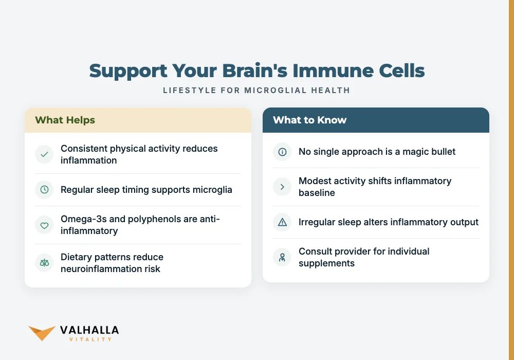
Evidence for lifestyle modulation of neuroinflammation is real and growing. No single approach achieves the precision of a targeted pharmacological intervention, but several factors reliably shift the microglial environment in a favorable direction when sustained consistently.
How lifestyle factors support microglia function
Physical activity carries the strongest evidence. Exercise reduces peripheral inflammatory markers and may directly influence microglial activation states. Compound resistance training consistently reduces systemic inflammation, which in turn reduces the peripheral signals that prime microglia toward chronic overactivation. Even modest, consistent activity shifts the inflammatory baseline over time.
Sleep has a direct mechanistic connection to microglial function through the circadian regulation documented by Kou et al. (2024). Consistent sleep timing and adequate duration are active components of brain health support, not merely recovery tools. The circadian sensitivity of microglia means irregular sleep schedules alter their inflammatory output before other effects of sleep disruption appear.
Diet shapes microglial behavior through multiple pathways. Omega-3 fatty acids, found in fatty fish, have anti-inflammatory properties relevant to microglia. Polyphenols in berries, olive oil, and leafy greens reduce oxidative stress that triggers microglial activation. Dietary patterns that concentrate these nutrients have the strongest observational associations with reduced neuroinflammation and lower long-term dementia risk. These are food-level nutritional observations. A licensed provider determines whether any individual supplementation is appropriate after reviewing labs and health history.
Stress reduction closes the loop. Chronic psychological stress elevates glucocorticoid levels, and prolonged glucocorticoid exposure shifts microglia toward a sensitized, pro-inflammatory phenotype. Consistent practices that reduce perceived stress interrupt this pathway regardless of the specific method.
The interaction between these factors is bidirectional. Poor sleep impairs stress resilience. Chronic stress disrupts sleep quality. A diet high in processed foods elevates the peripheral inflammatory markers that prime microglia. Addressing these factors together, rather than in isolation, produces more sustained effects on the microglial environment than any single change alone.
These strategies do not replace a clinical evaluation when neurological symptoms are present. They represent modifiable factors that, maintained over years, meaningfully influence the microglial environment and support both brain health support and long-term neurodegenerative disease prevention.
Take the next step
A licensed provider can review your full health history and help identify which factors may be most relevant to your long-term brain health. Book Your Consultation to connect with a provider.
FAQ
What do microglia do in the brain?
Microglia are the brain's primary immune cells, clearing cellular debris through phagocytosis, regulating inflammation through cytokine production, pruning synaptic connections, and monitoring the neural environment continuously. A 2025 paper by Hume in Trends in Neurosciences confirms that brain tissue without functioning microglia rapidly develops uncontrolled infection and progressive neurodegeneration.
What happens when microglia become overactive?
Overactive microglia release pro-inflammatory cytokines and reactive oxygen species continuously, damaging the neurons they are meant to protect. They also prune synapses indiscriminately, which disrupts memory and cognitive flexibility. This state, reactive microgliosis, is a central mechanism in Alzheimer's disease, Parkinson's disease, and other neurodegenerative conditions.
How does microglial dysfunction affect brain health?
Microglial dysfunction allows neurotoxic debris to accumulate, sustains chronic neuroinflammation, and disrupts the synaptic maintenance that supports memory and cognition. Research by Wang et al. (2021) shows these dysfunctional states can persist through epigenetic mechanisms even after the original trigger is removed, which is why early attention to neuroinflammatory risk factors matters more than late intervention.
What diseases are linked to dysfunctional microglia?
Alzheimer's disease, Parkinson's disease, multiple sclerosis, and ischemic stroke each involve significant microglial dysfunction. In Alzheimer's disease, microglia fail to clear amyloid aggregates and shift into states that amplify tau pathology. In Parkinson's disease, sustained microglial activation in the substantia nigra accelerates the loss of dopamine-producing neurons.
Can neuroinflammation be reduced naturally?
Regular physical activity, consistent sleep, an anti-inflammatory diet rich in omega-3 fatty acids and polyphenols, and chronic stress reduction each support healthier microglial function over time. These are long-term strategies, not acute treatments. Whether any specific supplement or clinical intervention is appropriate requires evaluation by a licensed provider.
Sources
- Hume DA (2025). Life without microglia. Trends in neurosciences. PubMed
- Kou L et al. (2024). Circadian regulation of microglia function: Potential targets for treatment of Parkinson's Disease. Ageing research reviews. PubMed
- Valiukas Z et al. (2025). Microglial activation states and their implications for Alzheimer's Disease. The journal of prevention of Alzheimer's disease. PubMed
- Shi FD et al. (2025). Neuroinflammation across neurological diseases. Science (New York, N.Y.). PubMed
- Planas AM (2018). Role of Immune Cells Migrating to the Ischemic Brain. Stroke. PubMed
- Wang L et al. (2021). Epigenetic Modulation of Microglia Function and Phenotypes in Neurodegenerative Diseases. Neural plasticity. PubMed
You May Also Like
 Featured
At Home STI Test: How It Works, Accuracy & Privacy
At home STI test kits let you screen for chlamydia, HIV, and more with private, rapid STI results. Learn how to use them and what to do next.
November 5, 2026 ETRead more → Featured
What Are Endorphins? Benefits, Signs, and How to Boost Them
What are endorphins? Learn how these brain chemicals affect pain relief, mood, and energy, plus signs of low endorphins and how to boost them.
October 25, 2026 ETRead more → Featured
Tirzepatide vs Semaglutide: The Complete Comparison
Tirzepatide vs semaglutide: compare mechanisms, effectiveness, side effects, and what to discuss with a licensed provider before starting either.
October 24, 2026 ETRead more →